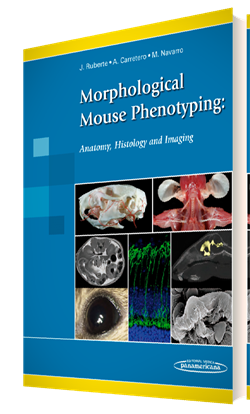
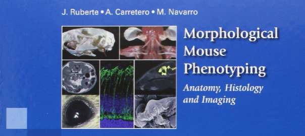
If you use mice as animal model in research projects, make sure not to miss this new atlas of mouse anatomy:

París, Jesús Ruberte, Romay Ana Carretero, and Beltrán Navarro. Morphological Mouse Phenotyping: Anatomy, Histology and Imaging. 1st ed. Editorial Médica Panamericana S.A., 2016.
From the back cover of the book: Animal models of disease are fundamental in research to improve human health. The success of using genetically engineered mice to evaluate molecular disease hypotheses has encouraged the development of massive European and global projects making the mouse the most used animal model. Consequently, laboratory mouse populations are straining the housing capacity of pharmaceutical and biotechnology companies, as well as public research institutions. However, the scientific community often lacks sufficient expertise in morphological phenotyping to effectively characterize and validate these animal models.
Although the mouse displays fundamental morphological similarities to humans, a mouse is not a man. Here we present a complete and integrative description of normal mouse morphology. The main characteristics of this book are:
- More than 2.200 original images have been specifically produced for this book in the Mouse Imaging Platform (Center for Animal Biotechnology and Gene Therapy, Universitat Autònoma de Barcelona).
- These images show the anatomy, histology and cellular structure of mouse organs.
- In addition, correlative X-ray, Computed Tomography, Magnetic Resonance (*) and Ultrasound images complete this integrative vision of the mouse morphology.
- Classical anatomical techniques such as conventional dissection, skeletal preparations, vascular injections, as well as histological, immunohistochemical and electron microscopy techniques have been employed to characterize the mouse morphology.
This book, essentially an atlas, also contains explanatory diagrams and text that guides the reader through normal mouse anatomy, histology and imaging, and is aimed for mouse researchers as well as veterinarian and human pathologists.
The book is available from the publisher, from Amazon.com (check also your country website if available), and from other places.
(*) all Magnetic Resonance images in this book were acquired by Dr. Silvia Lope-Piedrafita at the at the NMR service (SeRMN) of Universitat Autònoma de Barcelona, in a 7 Tesla Bruker BioSpec 70/30USR spectrometer. SeRMN has been an active partner of the jMRUI community since many years.
